MEA


The MEA set-up is used as a noninvasive method for monitoring spontaneous and evoked activity (spikes) as a change in the electrical potential of the intercellular space surrounding the cell. Spikes are a particular class of electrophysiological signals that depend on the excitably characteristics of cells and on their nature to generate Action Potentials (APs); APs base on the depolarization and repolarization of cell membranes due to ions flux (Ca+, K+ and Cl-).
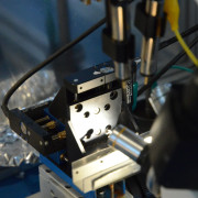
Every treatments that alter the ability of membrane to exchange ions reflect on the electrical activity of the cells, thus on the signals recordable. A typical setup for MEA recording is based on metal microelectrodes fabricated on a planar chip, discrete-element preamplifiers located close to the MEA device and a multi-wire cable that conducts the pre-amplified analog signals to a data acquisition card.
We have already reported (Masi et al. 2009, PNAS 106:4048-4053) time recordings of single-unit spike activity with MEAs in acute slice of Zea mays L. root apex. Extracellular electrical activity (spikes) can be recorded simultaneously from 60 electrodes (30 μm diameter) with high spatial and temporal resolution. The nature of spike shapes has been studied on each MEA electrodes (200 μm interelectrode spacing).
This new technique allows us to map functionally discrete regions of the root and to observe the space-time relationships and the spontaneous electrical activity of the root apex both in normal condition and in response to stress (drug treatment, gravistimulation, heat, electrical stimulation etc). Synchronous events and the propagation of signals can also be investigated.